A PEG-lipid makes up a small fraction of any lipid nanoparticles formulation. Its influence far exceeds its share. The PEG-lipid anchor structure determines how long the polymer layer survives on the particle surface. This question sits at the center of practical lnp design.
Anchor length and chemistry create a tunable trade-off. DSPE-PEG holds its position firmly. DMG-PEG 2000 releases quickly. Each choice shifts the balance between circulation stability and transfection performance.
This article connects structural features of peg lipids to real formulation decisions. It shows how peg-lipids behave in practice.
The short answer: the PEG-lipid anchor governs shedding kinetics, and shedding kinetics drive stability and biological fate.

Key Takeaways
-
PEG-lipid anchor structure controls how fast the polymer layer sheds from the particle surface.
-
Longer, saturated anchors like DSPE-PEG stay on the particle longer and improve circulation stability.
-
Shorter anchors like DMG-PEG 2000 shed quickly and can enhance transfection but may reduce storage stability.
-
PEG surface density must balance stealth against cellular uptake; moderate coverage often works best.
-
Choose PEG-lipids based on delivery goals and confirm performance with lab measurements.
PEG-Lipid Anchor Structure Basics
Anchor Length and Saturation
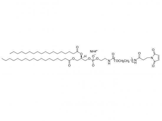
The lipid anchor structure begins with the hydrocarbon chain. DMG carries two C14 myristoyl chains. DSPE carries two C18 stearoyl chains. The extra four carbons per chain let DSPE-PEG insert more deeply into the particle membrane. Deeper insertion creates more van der Waals contacts with neighboring lipids.
Acyl chain length and saturation change membrane affinity in predictable ways. DMG-PEG 2000 anchors shallowly and exchanges rapidly with serum lipoproteins. DSPE-PEG holds its position and stabilizes the particle for longer. For related materials, see SINOPEG PEGylated Lipid products.
| Anchor | Insertion behavior | Stability outcome |
|---|---|---|
| DMG-PEG (C14) | Shallow anchoring, rapid dissociation | Size growth over time, falling particle counts |
| DSPE-PEG (C18) | Deep anchoring, strong retention | Size increase under 12 nm after 4 h, no concentration drop |
C14 anchors insert shallowly and exchange with serum lipoproteins, while C18 anchors insert deeply and stabilize durably.
Anchoring Chemistry and Linkage
The linkage between headgroup and anchor also matters. Amide, ether, and ester bonds differ in hydrolytic stability. Ester bonds can cleave; amide bonds resist breakdown. Saturated chains pack tightly and lower membrane fluidity around the anchor. Unsaturated chains pack loosely and weaken retention.
These features set the stage for shedding. Acyl chain saturation and acyl chain length together define how firmly peg lipids sit in the bilayer. Short, unsaturated lipid anchors leave quickly. Long, saturated lipid anchors stay. Formulators read this peg-lipid anchor structure as a retention dial before any bench work begins.
PEG Shedding and Surface Density
What Drives PEG Shedding
PEG-lipid shedding describes the loss of PEG molecules from the particle surface after formulation. Three mechanisms drive this process, and the peg-lipid anchor structure modulates each one.
-
Desorption: labile dimyristoyl-based peg lipids such as DMPE-PEG leave lipid nanoparticles rapidly in blood plasma. This depegylation precedes protein corona formation, including apoE adsorption onto depegylated particles.
-
Exchange with biological membranes: dimyristoyl-based peg lipids redistribute from particles to endogenous plasma components such as lipoproteins, which act as hosts. Net transfer of lipid-tailored entities to lipoproteins has been reported.
-
Dilution during formulation: this mechanism receives less attention in the literature, yet formulators should treat it as a design variable rather than an afterthought.
Anchor length sets the pace. Short anchors shed faster. Long, saturated lipid anchors resist shedding. DSPE-PEG generally associates more strongly with lipid assemblies and may remain in LNPs longer than DMG-PEG. Rapid peg shedding from DMG-PEG 2000 therefore reflects weak anchoring, not a formulation defect.
PEG Lipid Surface Density Effects
Surface coverage matters as much as anchor chemistry. Depending on chain length and peg grafting density, the hydrophilic peg chain arranges into a mushroom-like conformation at low coverage or a brush conformation at high coverage. A sparse mushroom-like conformation permits partial protein adsorption. A dense brush conformation extends chains outward and repels proteins more effectively.
| PEG surface density (chains/nm²) | Regime | Protein effect | Clearance outcome |
|---|---|---|---|
| Below 0.32 | Sparse coverage | Substantial serum protein adsorption, minimal shielding | Accelerated immune recognition |
| 0.32 to 0.96 | Intermediate | Partial shielding, some ligand accessibility | Moderate reduction in opsonization |
| Above 0.96 | Brush conformation | Near-complete suppression of nonspecific adsorption | Prolonged circulation |
Higher peg surface density reduces nonspecific adsorption but can impede functional ApoE recruitment. In one study, DMG-PEG2k at 1.5 mol% produced strong ApoE binding, while 5.0 mol% significantly reduced ApoE association. Regular PEGylation below 3 mol% keeps the lnp membrane accessible for receptor recruitment. Coverage above 3 mol% maximizes stealth at the cost of cellular interaction.
The hydrophilic PEG chain builds a flexible hydrated barrier around each particle. This layer shields carriers from aggregation, enzymatic attack, and nonspecific adsorption, and it supports lnp stability during storage and dilution. The hydrated barrier also limits recognition by the mononuclear phagocyte system. DSPE-PEG2000 illustrates the design: a hydrophobic anchor inserts into the bilayer while the PEG2000 chain extends outward.
Shedding kinetics feed directly into biological fate. Falling surface PEG density exposes the surface to opsonins and speeds clearance. Repeated dosing adds another layer, since anti-PEG IgM can trigger accelerated blood clearance, with complement activation peaking 3 to 4 days after the first dose. Balanced peg chain density and mixed chain lengths preserve lnp stability while retaining the surface recognition that hepatic uptake requires.
PEG Lipids, Retention, and LNP Membrane Stability
Stability Versus Transfection
Retention cuts both ways. A long-lived PEG layer supports colloidal stability and extended circulation. The same layer can block endosomal escape. The particle reaches the cell interior, yet the polymer coat still shields the membrane from fusion and disruption. Transfection efficiency falls.
Faster shedding reverses the pattern. DMG-PEG 2000 leaves the surface quickly, and the exposed bilayer interacts more readily with endosomal membranes. Cellular uptake and protein expression can improve. The cost appears in storage and in circulation, where particles may aggregate or clear early.
PEG lipids control particle size and prevent aggregation through a hydrophilic steric barrier. This barrier blocks serum-protein interactions and inter-particle contact. It also preserves particle integrity across formulation, storage, and administration. See SINOPEG lipid products for related lipid materials.
| Mechanism | How PEG Lipids Achieve It |
|---|---|
| Prevention of particle aggregation | Steric and electrostatic hindrance lowers the likelihood of aggregation in storage or in blood plasma |
| Prolonged circulation time | The PEG corona reduces plasma protein adsorption and slows clearance by the mononuclear phagocyte system |
| Improved formulation consistency | Uniform particle size and reproducibility across batches |
PEG-lipids are the primary lipid used to control aggregation as PEG creates a steric barrier to aggregation during storage. It is worth noting that PEG-lipids inhibit but not prohibit lipid nanoparticle aggregation.
Architecture adds another variable. Branched PEG-lipid conjugates confer higher stealth properties to lipid nanoparticles than linear PEGs. Branched structures therefore extend circulation beyond what a linear chain of similar size delivers.
Matching Retention to Application
Application context sets the target retention profile. Systemic delivery into the bloodstream rewards durable anchoring, because the particle must survive dilution and protein exposure before it reaches target tissue. Repeat dosing complicates the picture. Successive administrations can provoke anti-PEG responses, and a formulation that sheds gradually may reduce the accumulated polymer burden at the surface.
Tissue targeting pulls in the opposite direction. Hepatic uptake depends on ApoE adsorption onto the particle surface. An overly dense or overly persistent PEG layer suppresses that recruitment. Formulators here want moderate retention that leaves the lnp membrane accessible.
The peg-lipid anchor structure tunes this balance without a full reformulation. Swapping DSPE-PEG for DMG-PEG 2000 changes shedding kinetics while the ionizable lipid, helper lipid, and payload stay fixed. That single substitution shifts lnp stability, membrane fluidity at the anchor site, and the eventual biological fate. Branched or novel anchors widen the design space further, since they decouple stealth from chain length.
Membrane fluidity deserves attention during this work. Saturated C18 chains pack tightly and stiffen the local bilayer. Shorter chains leave the surrounding lipids more mobile. Both effects feed into lnp membrane stability and into how the particle behaves after shedding begins.
No single retention profile serves every program. The right choice follows from the delivery route, the dosing schedule, and the target tissue. Anchor chemistry gives formulators a practical lever for reaching that profile. For a specific example of a functionalized DSPE-PEG derivative, see DSPE-PEG-PBA.
Selecting and Screening PEG-Lipids
Choosing Based on Formulation Goals
Selection starts with the job the PEG-lipid must do. Formulators should define required surface retention, particle stability, cargo type, and delivery objective before comparing chain lengths. A structured sequence keeps the choice grounded.
-
Define what the PEG-lipid must accomplish for the program.
-
Match the anchor to the needed shedding profile.
-
Run a controlled head-to-head comparison when behavior is uncertain.
-
Measure rather than infer.
-
Decide on measured outcomes.
Holding cargo, ionizable lipid, helper lipid, cholesterol, and method constant makes comparisons fair. Matching molecular weight, such as DSPE-PEG2000 against DMG-PEG 2000, isolates the anchor variable. The table below summarizes the general tendencies.
| Criterion | DSPE-PEG | DMG-PEG |
|---|---|---|
| Anchor | Phospholipid, two C18 chains | Glycerol-based, two C14 chains |
| Anchoring strength | Generally stronger | Generally more readily released |
| Shedding profile | Often longer retention | Often faster shedding |
| Stability fit | Persistent coverage needed | Surface exposure intended |
These tendencies guide early choices for peg lipids, yet measurement confirms them. Composition shapes surface dynamics and in vitro performance, and anchor length tunes pharmacokinetics. That makes peg lipids central to lnp design and to any optimal lnp design strategy.
Key Parameters to Measure
Screening tracks physical and functional quality attributes together. Particle size, PDI, zeta potential, encapsulation efficiency, and RNA integrity belong on every batch record. PEG surface density over time reveals shedding behavior directly. Storage and dilution studies test lnp stability under real conditions, including long-term storage and accelerated conditions.
Zeta potential for PEGylated LNPs should target near-neutral values of -10 to +10 mV to minimize non-specific interactions while maintaining colloidal stability.
Functional potency follows encapsulation. Cell-based assays confirm protein expression for mRNA or target knockdown for siRNA. Tracking surface peg density alongside transfection links peg surface density to biological outcome. Suppliers such as SINOPEG offer PEG derivatives and PEGylated lipids at milligram to kilogram scale, one sourcing option among several. Structural predictions still require experimental confirmation, since measured retention and delivery performance remain the final arbiter.
The peg-lipid anchor structure sets the pace of shedding. Shedding then shapes particle stability, protein interactions, and transfection. DSPE-PEG holds its place and supports long circulation. DMG-PEG 2000 leaves quickly and favors cellular exposure.
No single peg lipid wins everywhere. Each program weighs stability against potency, and the delivery route decides which side matters more. Structural reasoning narrows the options, yet measured retention and potency remain the final test. Formulators should treat peg lipids as a design variable and confirm every choice at the bench.
FAQ
Which anchor length suits long circulation?
Long, saturated anchors resist shedding. DSPE-PEG carries two C18 chains and holds the surface layer longer. Short C14 anchors such as DMG-PEG 2000 leave quickly. Programs that need durable coverage usually start with the longer anchor.
Why do peg lipids shed from the particle surface?
Three mechanisms drive loss: desorption, exchange with plasma components, and dilution during formulation. Anchor structure sets the pace of each. Weak anchors release fast, and strong anchors resist. Formulators treat shedding as a design variable, not a defect.
Does a denser PEG layer always improve performance?
No. Dense coverage suppresses protein adsorption and extends circulation. It also blocks ApoE recruitment and endosomal escape. One study found 1.5 mol% DMG-PEG2k supported strong ApoE binding, while 5.0 mol% reduced it. Moderate coverage often serves delivery better.
How should a team screen peg lipids?
Hold the ionizable lipid, helper lipid, cholesterol, and payload constant. Swap only the anchor, matching molecular weight where possible. Track size, PDI, zeta potential, PEG density over time, storage stability, and transfection. Measured retention and potency decide the choice.