PEG-lipid selection balances four forces: molecular properties, application needs, regulatory requirements, and cost. Formulation scientists weigh each force when they choose PEG lipids for mRNA delivery systems.
Four factors drive the decision. PEG chain length and architecture set surface shielding and clearance. Lipid anchor and linker chemistry govern membrane retention and shedding. Application requirements shape dosing, targeting, and stability. Regulatory and cost considerations determine which options reach the clinic and the market.
This article offers a practical decision framework for peg lipid selection in lnp-mediated mrna delivery. Readers will learn how peg-lipid content, lipid nanoparticles design, and PEGylated lipid choice interact. No single option fits every program. A structured, iterative approach supports confident PEG-lipid selection.
Key Takeaways
-
PEG chain length and shape control how long the lipid nanoparticle stays in the body and how well it avoids immune clearance.
-
The lipid anchor and linker determine how firmly the PEG lipid stays on the particle and when it sheds, affecting stability and cell uptake.
-
Application needs like delivery route, dosing schedule, and storage guide which PEG lipid works best for your mRNA program.
-
Regulatory rules and cost factors limit which PEG lipids can reach the clinic and the market, so check these early.
-
No single PEG lipid fits every program; test candidates step by step to find the best match for your mRNA delivery system.
PEG Lipids in LNPs: Chain Length and Architecture
PEG lipids in LNPs form a dynamic steric brush at the particle surface. Chain length and architecture set the thickness of that brush. The brush, in turn, controls surface PEG exposure, particle stability and circulation time, and the route by which the body clears the particle. Formulation teams compare candidates along both axes before fixing a lead.
PEG Molecular Weight and Clearance
PEG chain length and molecular weight drive three linked outcomes: shielding, residence time, and clearance. Longer chains build a thicker hydration layer. That layer repels opsonins and slows renal filtration. The table below summarizes the relationships.
| Parameter | Surface shielding | Circulation time | Clearance |
|---|---|---|---|
| Chain length, density, terminal group | Tunable stealth behavior; brush thicker than immunoglobulin Fc domains and complement C3 convertases | Epitope shielding extends systemic circulation | Renal filtration or monocyte uptake outpaces chain rearrangement |
| Branching (Y-shaped, four-arm) | Concentrates handles for high-density brush layers | High-MW branched structures can sustain week-long circulation | Avoids viscosity of ultra-high-MW linear chains |
| Molecular weight | Larger hydrodynamic volume raises excluded volume | Higher MW extends residence time and flattens the concentration-time curve | Slows renal filtration |
Half-life rises with molecular weight. One published dataset shows a jump from about 18 minutes at 6 kDa to about 16.5 hours at 50 kDa. Clearance falls in parallel. PEG below 20 kDa leaves via urine, and elimination shifts from renal to hepatic as PEG mass grows. PEG lipids in LNPs sit at only about 1.5% of the formulation, yet they prevent aggregation during storage and shield lipid nanoparticles from immune clearance. Peg-lipid content stays low in most formulations. Branched structures reach high molecular weight without the viscosity penalty of ultra-high-molecular-weight linear chains.

Chain length carries an immunologic caveat.
PEG chain length is a critical structural determinant of immunological safety. The relationship is biphasic: both long-chain and short-chain PEG conjugates are more prone to triggering the ABC phenomenon.
Intermediate chains often balance shielding against this risk. Terminal chemistry is a separate design lever. Methoxy-capped PEG lipids suit non-reactive stabilization. Maleimide couples to thiols for ligand display. NHS esters bind amines. Azide, DBCO, and alkyne groups enable click workflows. Post-insertion of a pre-made ligand–PEG–lipid conjugate offers a modular route for rapid comparative screening. Each group must match the intended conjugation chemistry, and its stability in the formulation must be verified.
Linear Versus Branched PEG
Both linear and branched PEG chains can improve targeting behavior and transfection in mRNA delivery. Architecture is a fit question, not a quality ranking. Branched and Y-shaped PEG can markedly lower anti-PEG antibodies compared with linear PEG, which matters for repeated dosing. Branched forms also pack multiple handles at one point, so they build dense brush layers. Linear PEG lipids remain simpler to source, characterize, and insert. Programs with single administration often find linear chains sufficient. Programs facing repeat dosing may prefer branched architectures to manage anti-PEG antibodies.
These molecular choices feed directly into peg-lipid selection. PEG chain length and molar ratio together set surface density, and surface density shapes both shielding and ligand reach. A higher peg-lipid content thickens the brush but can limit ligand accessibility. A lower peg-lipid content favors interaction with target cells. Peg-lipid content and architecture interact inside the same particle. The peg-lipid composition of the particle follows from these choices. Formulators often treat peg optimization as an iterative loop: adjust chain length, adjust the ratio, then measure mRNA expression. PEG lipids in LNPs therefore reward systematic comparison across candidate structures for any mRNA delivery program.
Anchor and Linker Chemistry for PEG-Lipid Selection
The lipid anchor and the linker sit beneath the PEG chain. Together they decide how long a PEG lipid stays on the particle. Anchor structure sets membrane retention. Linker chemistry sets shedding. Both parameters feed directly into peg-lipid selection for any mRNA program.
Lipid Anchor and Membrane Retention
Anchor tail length controls how firmly a PEGylated lipid holds its place in the bilayer. A published in vivo dataset measured desorption one hour after administration:
| PEG-lipid alkyl/acyl chain length | Desorption at 1 h post in vivo administration | Retention implication |
|---|---|---|
| C14 dialkyl | 45% | Moderate dissociation; sufficient for self-assembly, prone to dissociation near lipid sinks |
| C16 dialkyl | 1.3% | Strong retention; improved LNP stability in biological fluids |
| C18 dialkyl | 0.2% | Strongest retention; minimal desorption, prolonged circulation |

Longer tails reduce desorption. Short C12 and C14 chains still support self-assembly, yet they dissociate readily into plasma, lipoproteins, and cell membranes. DSPE-PEG carries a C18 saturated dual acyl anchor and generally shows more sustained PEG retention than DMG-PEG, which carries a C14 dual acyl anchor. Each anchor fits a different job. C14 anchors suit programs that want rapid surface remodeling after administration. C18 anchors suit programs that need durable shielding.
Retention carries a trade-off. Persistent peg lipids improve particle stability and circulation time and support accumulation outside the liver. The same persistence limits cellular interaction and endosomal escape, so transfection can fall and dose requirements can rise. Peg-lipid content and tail length therefore move together. A higher peg-lipid content with long tails maximizes stealth. A lower peg-lipid content with shorter tails favors cell engagement.
Linker Chemistry and Shedding
The linker connects anchor to chain, and its stability profile sets the shedding rate. Stable linkages keep the PEG brush intact through storage and circulation. Cleavable linkages let the chain leave the surface at a chosen point, which can restore interactions between lipid nanoparticles and target cells. Neither route is superior in general. Stable linkers fit applications that demand long circulation and a fixed surface. Cleavable linkers fit applications that need a stealth phase followed by active uptake, a common pattern in mRNA delivery.
Linker choice also affects chemical stability during storage. Formulators verify that the linker survives the formulation pH and buffer and that it does not degrade into reactive species. A linker that hydrolyzes early leaves the particle under-shielded before it reaches the target tissue. Peg lipids in LNPs sit at low molar fractions, so a small loss of surface coverage can shift particle behavior. Analytical tracking of intact PEG lipid across time points gives an early signal of shedding.
Anchor and linker decisions rarely stand alone. They interact with chain length, with peg-lipid content, and with the administration route. Teams that screen anchors and linkers as a matrix resolve these interactions faster. That matrix approach keeps peg lipid selection grounded in measured retention and measured transfection for each mRNA delivery route.
Peg lipids in LNPs also shape storage behavior. Peg lipids with stable anchors protect lipid nanoparticles against aggregation, and that protection supports shelf life. Short-chain peg lipids shed quickly, so they fit single-dose programs that need fast surface remodeling. A formulation team can compare candidate peg lipids on retention, shedding, and transfection in one screen. Each mRNA program then selects the anchor and linker pair that matches its dosing plan.
Application Requirements in mRNA Delivery
Targeting, Dosing, and LNP Formulation
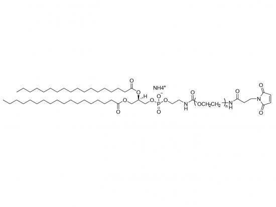
Delivery route, target tissue, ligand use, and cargo shape peg-lipid selection. Passive lymph-node targeting for vaccines relies on small, negatively charged particles near 30–50 nm. Passive tumor targeting exploits leaky vasculature. Immune-cell uptake improves with higher DMG-PEG2000 levels. Active ligand-mediated targeting uses DSPE-PEG2000-maleimide as a surface linker for mannose, anti-CD4, or PECAM-1 antibodies. Overcoming liver tropism calls for stealth strategies that prolong circulation.
| Targeting context | PEG-lipid choice | Effect on delivery |
|---|---|---|
| Passive lymph-node targeting | Tune peg-lipid content for ~30–50 nm size | Improved dendritic cell uptake |
| Immune-cell uptake | Increase DMG-PEG2000 concentration | Precise size control |
| Active ligand targeting | DSPE-PEG2000-maleimide linker | Cell-specific delivery |
| Overcoming liver tropism | Stealth PEG strategies | Broader biodistribution |
Ligands cannot compensate for poor size control or weak endosomal escape. Peg-lipid molar ratio sets surface density, and surface density governs both shielding and ligand reach. Frequent repeated dosing raises anti-PEG antibody production and accelerated blood clearance risk. Extending intervals or moderating peg density mitigates it. Moderate surface density preserves lnp stability while lowering immune recognition. Rational pegylation tuning balances these effects for repeated and targeted dosing in lnp-mediated mrna delivery.
Stability and Storage Needs
Formulation pH and buffer composition influence mRNA-LNP stability. Tris buffers outperform PBS, which causes acidic pH drift during freezing. A staged pH strategy holds ~4.0 during encapsulation, then shifts rapidly to ~7.4 for storage. Peg-lipid content near 1.5% and chain length govern steric stabilization during freeze-drying. Peg lipids in LNPs must match the lyophilization strategy rather than stay fixed.
Quality attributes guide peg optimization in every lnp formulation. Particle size and surface charge, mRNA encapsulation above 90%, lipid oxidation, and transfection efficiency form the core panel. Raising peg-lipid concentration shrinks particles and improves storage stability, yet excessive peg density suppresses uptake and endosomal release. A 1.5 molar ratio balances size and potency for intramuscular mrna delivery. Higher levels suit intravenous mrna delivery. Each lnp formulation design must resolve this trade-off through matrix screening.
Regulatory and Cost Considerations
Regulatory Compliance and Documentation
Regulators judge a PEG-lipid on safety, purity, and the paperwork behind it. Each requirement narrows the list of acceptable candidates before a formulation team ever runs a transfection assay.
| Requirement Category | Specific Regulatory Expectations for PEG Lipids |
|---|---|
| Purity and characterization | Specifications for purity, identity, and consistency; physicochemical characterization of lipid ratios, phase transition temperatures, surface charge, and morphology |
| Safety assessment | Cytotoxicity, genotoxicity, and immunostimulatory evaluation, including degradation products |
| Immunogenicity | Anti-PEG antibody monitoring, cytokine release profiling, and complement activation assessment |
| Impurity profiling | Residual solvents, heavy metals, and process-related impurities; ICH Q3 provides the general framework while LNP-specific criteria keep evolving |
| Documentation (CMC) | Detailed CMC packages, Quality by Design with defined critical quality attributes, validated analytical methods, and stability data |
Analytical workflows support these filings. Teams measure particle size and polydispersity by dynamic light scattering, check surface charge by zeta potential, and quantify encapsulated mRNA by encapsulation efficiency assays. Gel electrophoresis confirms mRNA integrity, and HPLC with mass spectrometry validates lipid purity. Stability testing across storage conditions then establishes shelf life. Suppliers with mature quality systems, such as Sinopeg with its ISO-9001 and ISO-13485 certifications, ease this documentation burden for peg lipids in an mRNA LNP program.
Cost and Supply at Scale
Raw materials rank among the key cost drivers in LNP manufacturing, alongside capital investment in mixing, microfluidics, and purification, plus consumables and scale-up yield losses. Techno-economic modeling places much of the operating expense in drug substance and drug product stages, driven by raw materials and downstream purification. LNP-formulated mRNA carries a cost premium of roughly USD 1.05 per dose against about USD 0.38 for adenoviral and USD 0.40 for saRNA formats. In silico Bayesian design-of-experiments modeling can predict operating cost reductions of 25–40% by identifying scalable encapsulation methods and optimal lipid ratios, including the PEGylated lipid share.
Supply strategy matters as much as unit price. Scale-up from preclinical to commercial volumes imposes lipid purity and batch-consistency requirements that compress the qualified supplier base. Clinical-stage growth forces formalized long-term supply agreements with GMP-qualified manufacturers. Dual-sourcing programs mitigate concentration risk. Lot-to-lot consistency of the lipid kit must be validated for transfer to GMP manufacturing. Peg-lipid content and overall lnp formulation choices therefore carry commercial consequences, and teams should confirm supply capacity before locking a lead.

A practical decision framework starts with application requirements. Teams define the route, dose schedule, and storage plan first. They then narrow candidates by molecular properties, including chain length, anchor, and linker. Regulatory and cost fit confirm the final choice.
No single PEGylated lipid is universally best. Each option fits different applications, and peg-lipid content must match the program. Formulation scientists should treat peg lipid selection as an iterative, application-driven process. Repeated screening across peg lipids, lipid nanoparticles, and mRNA cargo builds confidence. This approach supports reliable peg-lipid selection for lnp-mediated mrna delivery and keeps every mRNA delivery program on a defensible path.
FAQ
What drives PEG-lipid selection for mRNA delivery?
Selection starts with application needs. Route, dose schedule, and storage plan narrow the field. Molecular properties then refine the choice: chain length, anchor, and linker. Regulatory and cost fit confirm the lead. No single option suits every mRNA delivery program, so teams screen candidates iteratively.
How does PEG-lipid content affect particle performance?
PEG-lipid content near 1.5 molar percent balances size and potency for intramuscular mRNA delivery. Higher levels shrink particles and aid storage stability but suppress uptake. Lower levels favor cell engagement. Teams tune this ratio for each mRNA delivery route through matrix screening.
Which anchor suits long-circulating mRNA delivery?
C18 anchors retain strongly, with about 0.2% desorption at one hour in vivo. C16 anchors retain nearly as well at about 1.3%. Both support durable shielding for mRNA delivery. C14 anchors desorb faster and suit programs that want rapid surface remodeling after dosing.
Do branched PEG chains help repeated mRNA delivery?
Branched and Y-shaped chains can lower anti-PEG antibodies compared with linear chains. That matters for repeated mRNA delivery, where accelerated blood clearance poses a risk. Linear chains remain simpler to source and often suffice for single-dose mRNA delivery.
Which quality attributes guide optimization?
Teams track particle size, surface charge, mRNA encapsulation above 90%, lipid oxidation, and transfection efficiency. Buffer choice matters too: Tris outperforms PBS during freezing. These attributes let formulators compare a PEGylated lipid candidate across mRNA LNP batches and confirm the choice before scale-up.